Office
Complete Anatomy 11.5
OfficeUpdated
| Requirement | On file |
|---|---|
| macOS | macOS 11 or later |
| CPU | Apple Silicon, Intel 64-bit |
| Download | 838 MB |
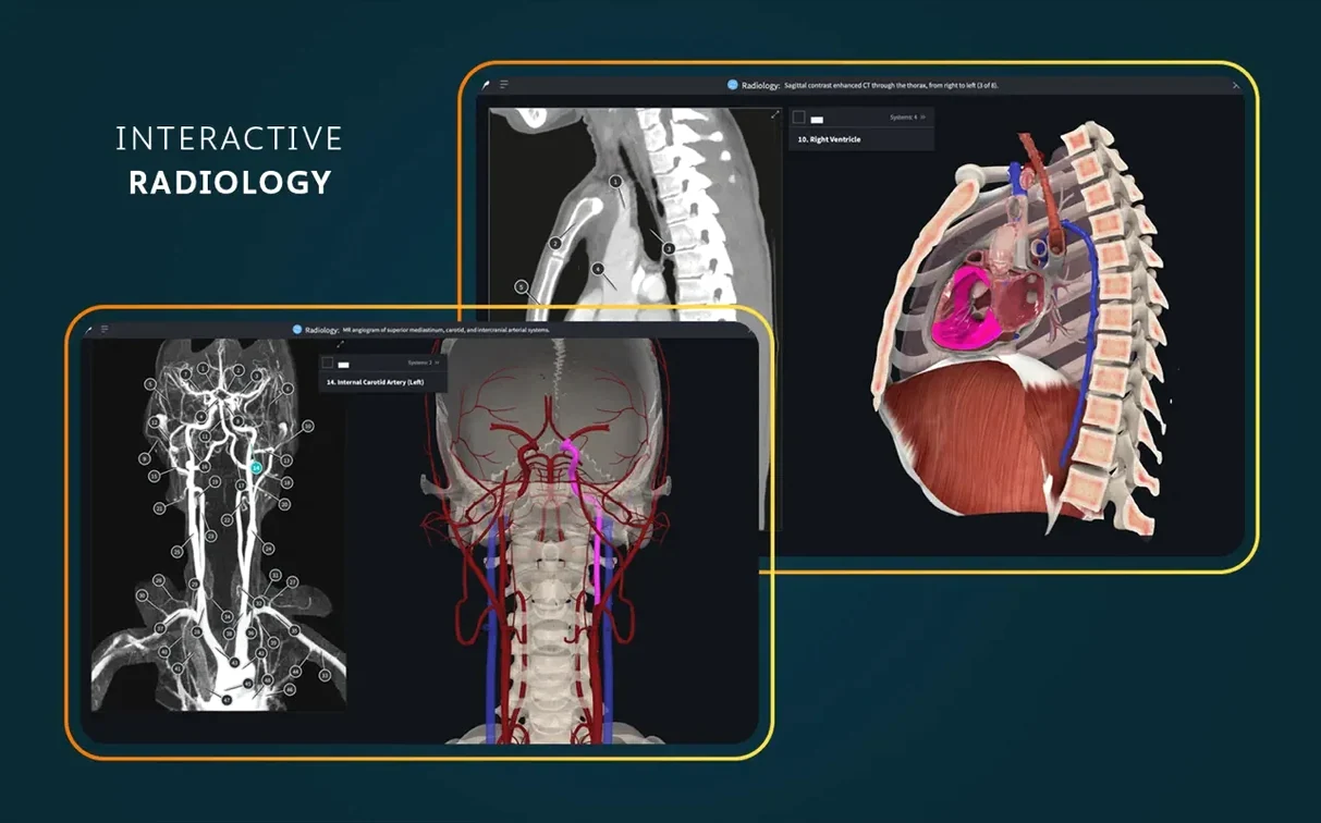
A single radiology view, placed beside an interactive 3D model, often reveals more than a dozen textbook plates. Complete Anatomy builds its macOS experience around that pairing, letting you trace a vessel on a CT slice while the volumetric model updates in real time.
Correlating cross-sections with radiology
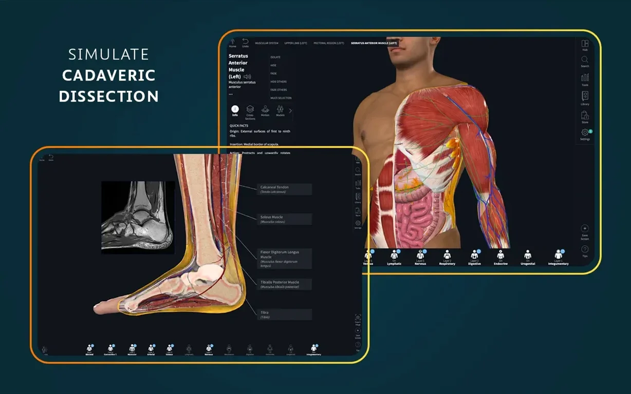
Dynamic cross-sections move in three axes, and the built-in radiology view maps the same plane onto a clinical-style image. You drag a section line and watch both representations shift together — useful when you need to connect gross anatomy with what appears on a scan.
Testing function through muscle movement
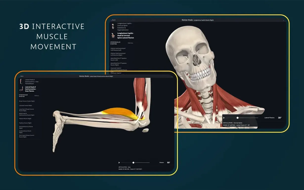
Real-time muscle movement shows origin, insertion and innervation at the same time. You isolate a muscle group, trigger the action and watch it play out on the model, pausing mid-contraction to check how neighbouring structures relate.
Working across architectures
The 11.5 release runs natively on ARM and x86 64-bit Macs from macOS 11 onwards, so switching between an Apple Silicon desktop and an older Intel machine does not change tool behaviour or model resolution.
Sign In, Then Strip Back to the Skeleton
You launch Complete Anatomy from the Applications folder or Launchpad. A sign-in screen appears — the app needs an account before it loads the full model library. Once you are in, the main screen defaults to the male skeletal system, fully articulated, suspended in the centre of the workspace.
You start by picking a region. Tapping the Systems menu in the top-left toolbar, you toggle off the skeleton and switch on the cardiovascular system. The heart appears, beating at a resting 72 bpm. You pause the motion with the spacebar, then double-tap the left ventricle to isolate it. The surrounding myocardium fades to 30% opacity, and a small info panel lists its wall thickness, blood supply and innervation.
Next you add a cross-section plane. Dragging the axial slider from the right-hand tools panel, you slice through the thorax at T4–T5. The cut surface reveals the four chambers in a flat anatomical plane, colour-coded by tissue type. You switch to the radiology tab, and the same plane now shows a corresponding CT slice. The correlation is immediate — the 3D model on the left, the scan on the right, both locked to the same coordinate.
You save the view as a Screen using the camera icon, give it a label (“Aortic valve — axial”), and drop it into a new folder for later review. Before closing, you open the Courses panel, pick a dissection module on the upper limb, and scrub through the first three steps. Each one adds or removes a layer of tissue, narrated by a short text block. The whole session, from launch to saving a custom view, takes under eight minutes.
A physiotherapy student preparing for a shoulder exam
Maria is in her third year of a physiotherapy degree. She has a practical exam on the rotator cuff in two days, and the 2D plates in her atlas are not helping her visualise the way supraspinatus tucks under the acromion during abduction.
She opens Complete Anatomy and loads the muscular system with the skeleton kept visible at 40% opacity. She searches for “supraspinatus” in the model browser, and the app centres the view on the right scapula, highlighting the muscle in red. She triggers the movement tool and selects shoulder abduction. The humerus rises, and she watches the supraspinatus shorten while the deltoid takes over mid-range. She pauses at 90 degrees and rotates the model 15 degrees posteriorly — now she can see the subacromial space narrow in real time.
She switches to the innervation overlay. The suprascapular nerve lights up in yellow, tracing back to the upper trunk of the brachial plexus. She screenshots the view, pastes it into her revision notes, and adds a text annotation: “Impingement zone at 60–120° — supraspinatus + subacromial bursa.”
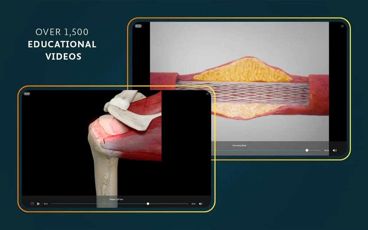
Before closing, she opens one of the 1,500 clinical videos, filtering by “shoulder” and “ultrasound.” A two-minute clip shows a dynamic ultrasound of the cuff during abduction, with the probe position mapped onto a 3D shoulder model in the corner. She watches it twice, then quizzes herself with the label tool: she hides all muscle names and tries to identify the four cuff tendons by shape and position alone. She gets three out of four — teres minor catches her out — so she bookmarks the muscle for review the next morning.
Break the Post-Update Login Loop
Sign-in loop after updating. Some users find that version 11.5 asks for credentials again even though they were signed in before the update. The fix is straightforward: quit the app completely (Command-Q, not just closing the window), delete the cached session file at ~/Library/Application Support/Complete Anatomy/, then relaunch and sign in fresh. The account data itself lives server-side, so no saved Screens or folders are lost.
Model rendering lags on older Intel machines. If the 3D view stutters during cross-section dragging or muscle movement, drop the texture resolution in Preferences > Graphics. Setting it to Medium keeps the radiology overlay crisp while reducing the GPU load. On a 2018 MacBook Air, this brought frame rates from roughly 12 fps back to a smooth 30 fps during real-time movements.
AR mode camera permission denied. The AR feature needs camera access, but macOS may block it silently after installation. Check System Settings > Privacy & Security > Camera and toggle the switch for Complete Anatomy. If the app was not listed, launch AR mode once, decline the prompt, then check again — the toggle will appear and you can enable it manually.
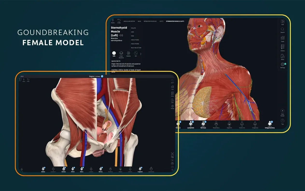
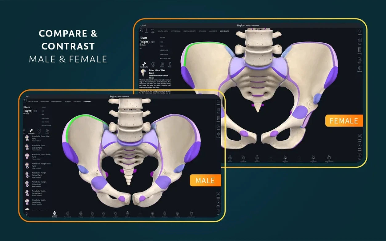
Female pelvis model not loading. The full female anatomy content requires an explicit download from the Content Library. Tap the cloud icon next to “Female Pelvis — Full” in the Regions panel and wait for the progress bar to finish. The model is roughly 180 MB, so a spotty Wi‑Fi connection can cause it to stall at 90%. Switching to a wired connection or downloading it before a study session avoids the frustration.
Search not returning expected results. The model browser uses English anatomical terms by default. If you are working with the interface set to German, Spanish, French or Simplified Chinese, the UI labels switch but the search index remains English-only. Typing “Musculus supraspinatus” finds nothing; “supraspinatus” finds the muscle instantly. Keep search queries in English regardless of the interface language.
Double-Tap to Isolate Any Structure
- Double-tap to isolate. A double-tap on any structure fades everything else to 30% opacity. Double-tap again to restore the full model. This works on bones, muscles, vessels and nerves — faster than hunting through the Systems menu.
- Spacebar toggles the heartbeat. When the cardiovascular system is active, the spacebar pauses and resumes the cardiac cycle. The heart rate slider at the bottom of the screen adjusts from 40 to 120 bpm, and the paused state holds at the exact phase of the cycle you stop at.
- Option-drag duplicates a Screen. In the Screens panel, holding Option while dragging a saved view creates a copy. Useful for making variations — same angle, different layer visibility — without rebuilding from scratch.
- Right-click for the quick menu. A right-click on any structure opens a radial menu with three options: Add Label, Hide, and Focus. “Focus” zooms and centres the view on that structure in one motion, bypassing the trackpad pinch-and-drag dance.
- Number keys switch saved tool views. If you save frequently used tool configurations (cross-section plane at T10, muscle movement set to knee flexion, radiology overlay enabled), you can assign them to number keys 1–9 via the Screens panel. Tapping “3” mid-study jumps straight to that setup.
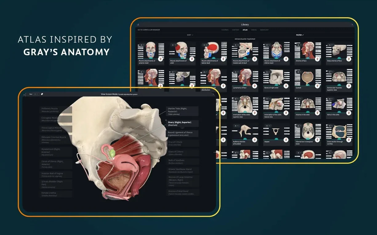
- Two-finger swipe browses the Atlas. In the Atlas view, a two-finger horizontal swipe on the trackpad flips through the 700+ plates without clicking the arrows. The same gesture works in Courses to move between dissection steps.
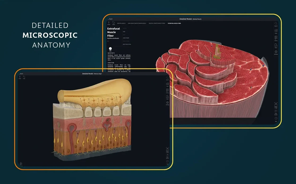
- Microscopic models have their own zoom behaviour. The 39 histology models (nephron, osteon, sarcomere and others) respond to scroll-wheel zoom differently from the gross anatomy models. Scrolling inward pushes past the outer membrane into the intracellular detail, and a small breadcrumb trail at the top shows your depth level — easy to miss if you are used to the standard zoom limits of the skeletal model.
- iOS 26 support — Optimized for the latest operating system version.
- Operating system: macOS 11.0 or later
- Architecture: Apple Silicon (ARM) and Intel (x86 64-bit)
- Account: A signed-in account is required for full functionality
Questions on file
Does Complete Anatomy 11.5 run on older Intel Macs?
Yes, the app supports both ARM (Apple Silicon) and x86 64-bit architectures and requires macOS 11.0 or later.
What imaging tools are included beyond the 3D model?
It includes dynamic cross-sections that move in three axes and a radiology view that correlates clinical-style slices with the interactive model.
Sign in · Create an account to add a field note.
No field notes yet. A measurement, a version, a machine — they appear after moderation.